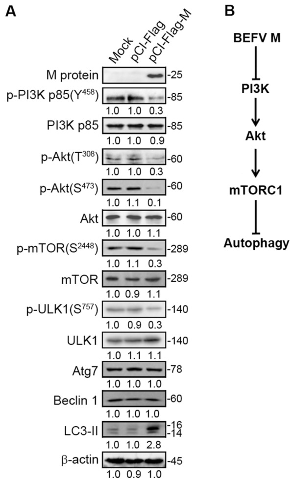
Figure 8.

The BEFV M protein induces autophagy through suppression of the PI3K/Akt/mTOR pathway. A MDBK cells were transfected with pCI-Flag-M and the cell lysates were subjected to immunoblotting with the indicated antibodies. The levels of the indicated proteins in the mock control were considered onefold. The protein levels were normalised to those for β-actin. The activation and inactivation folds indicated below each lane were normalized against values for the mock control. Signals were quantified using Image J software. The predicted size of each protein was labeled at the right-hand side in kDa. B A model illustrating BEFV M protein-induced autophagy through suppression of the PI3K/Akt/mTOR pathway.
