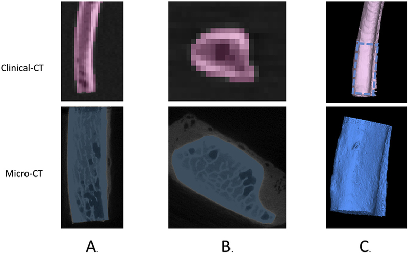
Fig. 2.
Rib specimen segmentation from the 0.50 mm slice thickness clinical-CT (pink) and micro-CT (blue) scans showing the sagittal view (A), axial view (B), and 3-D reconstruction of the micro-CT (blue) and clinical-CT (pink) with the region scanned with both modalities designated by dashed blue lines (C).

