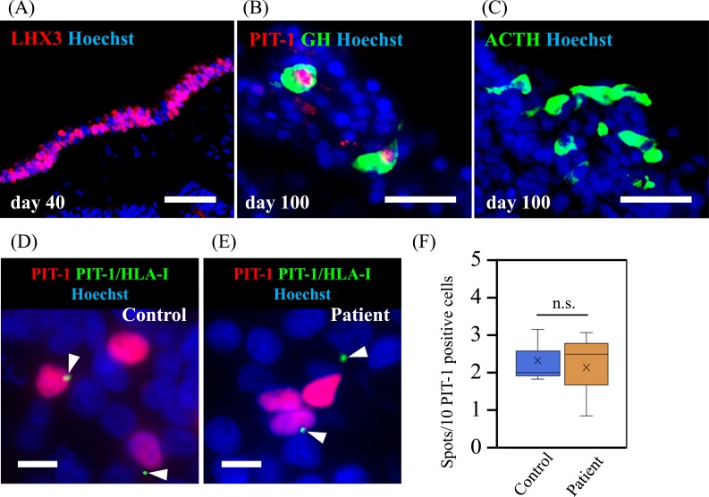
Figure 4.
PLA in human pituitary cells differentiated from iPSCs showed the presentation of PIT-1 epitope by MHC class I. iPSCs derived from the patient with anti–PIT-1 antibody syndrome and from a control subject were induced for the differentiation into anterior pituitary cells. (A) LHX3 expression on day 40, indicating the differentiation into oral ectoderm, which is a precursor of anterior pituitary cells. (B) PIT-1 and GH expression and (C) ACTH expression on day 100. Scale bar, 50 µm. PLA showed the presentation of PIT-1 epitope by MHC class I molecules in the anterior pituitary cells derived from the patient (D) and the control subject (E). Scale bar, 20 µm. (F) Quantitative analysis showed a comparable number in the presentation between the patient and the control subject. Three technical replicates were performed. Kruskal-Wallis test was used.

