
Fig. 3.
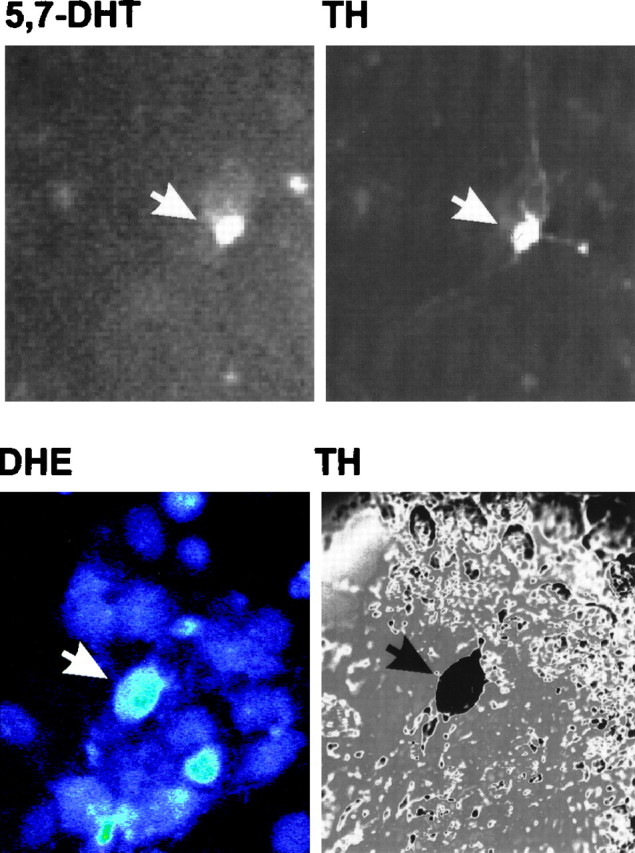
Identification of dopaminergic neurons using 5,7-DHT prelabeling or post hoc TH staining. Top panels, Mesencephalic cultures were incubated with 5,7-DHT for 30 min at 37°C, rinsed, and imaged by fluorescence microscopy with a computer-controlled camera. Images were taken at 40× magnification. Cultures were subsequently fixed and stained for TH using a CY3-coupled secondary antibody. Field relocation shows colocalization of TH with 5,7-DHT (white arrows). This methodology was used to assay changes in DHR and Rh 123 fluorescence in dopaminergic cells.Bottom panels, To measure DHE fluorescence in dopaminergic neurons, cultures were stained for TH via a color reaction after incubation with DHE. Bottom left panel, Confocal image of cells exposed to 6-OHDA for 30 min and incubated with DHE for 15 min (60×). On the right is the corresponding differential interference contrast image of TH-stained neurons in a relocated field.