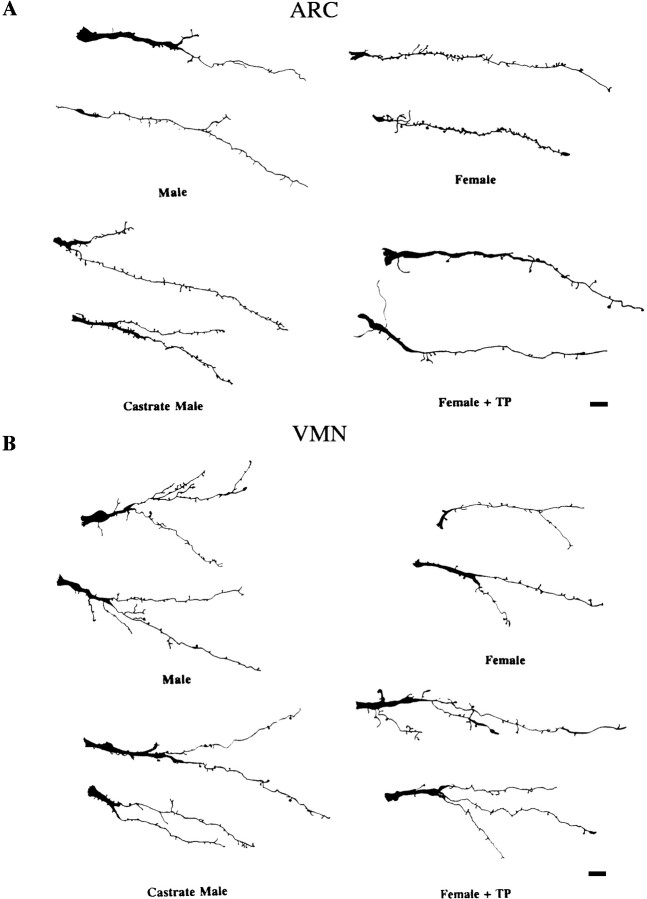
Fig. 3.
Camera lucida drawings of ARC andVMN dendrites from the hormonally treated groups. Drawings represent individual primary dendrites impregnated with Golgi-Cox from the ARC (A) andVMN (B) across four treatment groups. ARC dendrites from the groups with high levels of circulating testosterone (males and females + TP) have significantly fewer spines on their dendrites than do groups with low levels of testosterone (females and castrate males). Dendrites in the VMN have significantly more branch points in groups with high levels of circulating testosterone than do the groups with lower levels. Scale bars, 10 μm.

