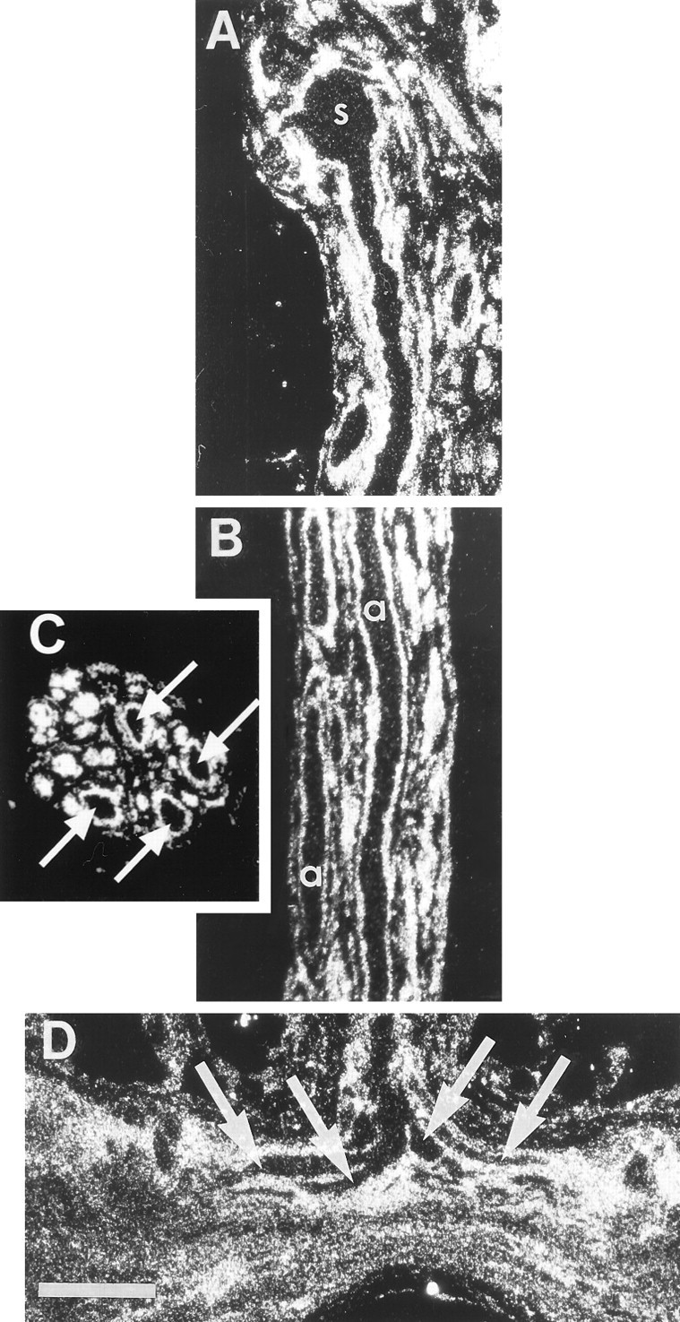
Fig. 1.

Glia, not photoreceptors, preferentially accumulate [3H]histidine. Dark-field autoradiographs of sections through preparations that had been incubated in 20 μm [3H]histidine in flashing light at 15°C. Silver grains over the glia outline the sparsely labeled photoreceptor profiles in each panel.A, Horizontal section through a photoreceptor soma (s) in the median ocellus and the initial portion of its axon in the median ocellar nerve. B, Horizontal section through the median ocellar nerve passes through two of the four photoreceptor axons (a). C, A cross section through the nerve shows the profiles of the four large, sparsely labeled photoreceptor axons (arrows) ringed by intensely labeled glia; other axons in the nerve are too small to be distinguished from their surrounding glia in cross section.D, Horizontal section through the commissure of the supraesophageal ganglion at the midline where the bifurcating primary branches of the presynaptic arbor (arrows) enter. Scale bar: D, 100 μm (applies to all panels).
