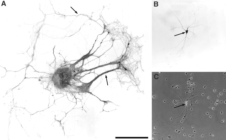
Fig. 1.
A, An OE explant from E14 rat maintained in vitro for 5 d extends numerous long axons from the explant onto the substrate. These axon bundles and processes strongly express the olfactory marker protein (arrows). B, A high-power micrograph showing an OB neuron expressing tyrosine hydroxylase after 10 d in culture (arrow). These neurons form extensive neurite arbors and contact other bulb cells within the culture.C, Phase-contrast micrograph of Bdemonstrating the presence of the TH-expressing neuron (arrow) and other OB neurons not expressing tyrosine hydroxylase. Scale bars: A, 1 mm (represents 150 μm inB and C).

