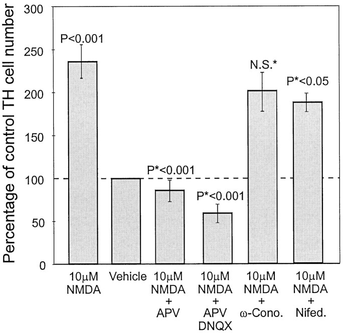
Fig. 5.
Quantification of TH-positive cells in cultures of NMDA-stimulated dissociated OB neurons treated with 100 μm APV, 100 μm APV and 10 μmDNQX, 500 nm ω-conotoxin GVIA, or 10 μmnifedipine. Control cultures all received sham pulses consisting of culture media. Pulses of NMDA at 10 μm increased the number of TH-positive OB neurons (p < 0.001), which were inhibited by APV (*p <0.001 vs NMDA-stimulated cultures), and APV–DNQX mixtures (*p<0.0001 vs NMDA-stimulated cultures). ω-Conotoxin did not significantly affect TH expression after NMDA stimulation; however, nifedipine reduced NMDA induction of TH from 143% increase to only 93% increase (35% decrease; *p < 0.05 vs NMDA-stimulated cultures).

