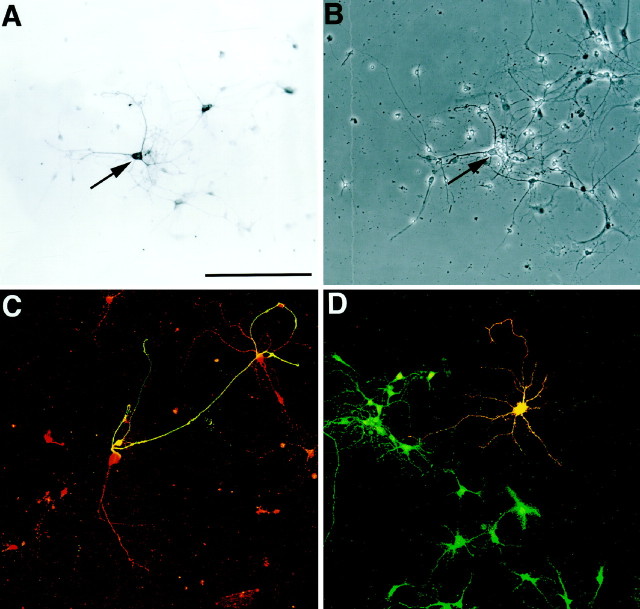
Fig. 7.
Expression of NMDA receptor subunits R1 and R2B in cultures of dissociated OB cells. A, NMDA receptor subunit R1 is expressed by many cells within the OB culture (arrow). These cells extend numerous fine processes and are often in contact with other cells in the culture. B, Phase contrast demonstrates that there are other cells within the cultures that do not express the NMDAR1 subunit. Some of these cells have the flattened appearance typical of glial cells in culture.C, Double immunofluorescence for NMDA receptor subunit R1 (red) and TH (green) shows that the neuron expressing TH also expresses this NMDA receptor subunit (yellow). D, Immunofluorescence for the NMDA receptor subunit R2A (green) and TH (red) demonstrates that the TH neurons in vitro also coexpress the R2A subunit. TH is distributed evenly throughout the neuron; however, the NMDA receptor subunit is localized to punctate deposits along the cell processes and over the cell perikarya. Processes from TH neurons often contain intense punctate deposits of NMDA receptor at process terminals. Scale bar:A, 120 μm (applies toA–D).

