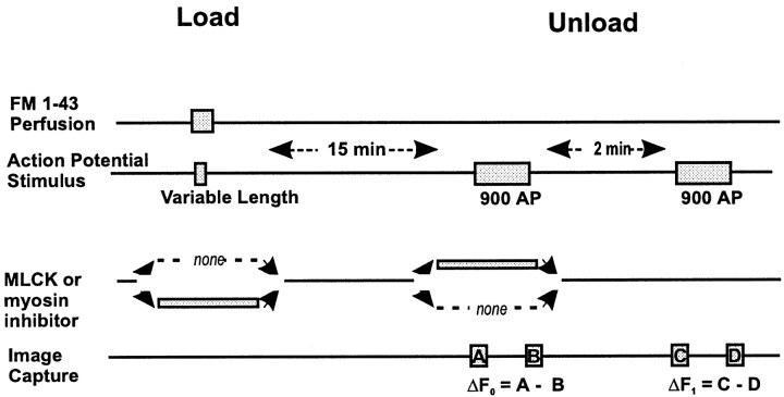
Fig. 1.
Protocols for FM1-43 labeling of vesicle recycling in the presence of MLCK or myosin inhibitors. Recycling vesicles were labeled by exposure to 15 μm extracellular FM1-43 (Load) during AP stimulus. The 90 sec of 10 Hz stimulation in a dye-free solution then released ∼90% of the vesicular dye in control conditions (Unload; ΔF0). A second round of 900 AP released the remaining 10% vesicular fluorescence (Unload; ΔF1). The impact of inhibitors of MLCK or myosin on vesicle recycling was determined by application during either the loading or the unloading phase. In either case, inhibitors were perfused in 150 sec before the stimulus and washed out after the stimulus.

