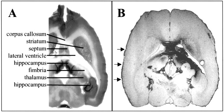
Fig. 6.
Immunohistochemistry for anti-hIgG Fcγ. A, Portion of Nissl-stained horizontal section at approximate level of B. Selected structures are labeled. B, Horizontal section from an animal infused with hIgG showing the typical distribution of immunoreactivity (right side, infusion site). In this animal, note the immunoreactivity in the septum bilaterally, the corpus callosum, the right striatum, the righthippocampus, and the fimbria and light immunoreactivity in theleft hippocampus and the fimbria. Arrowsmark the immunoreactivity in pia surrounding the brain.

