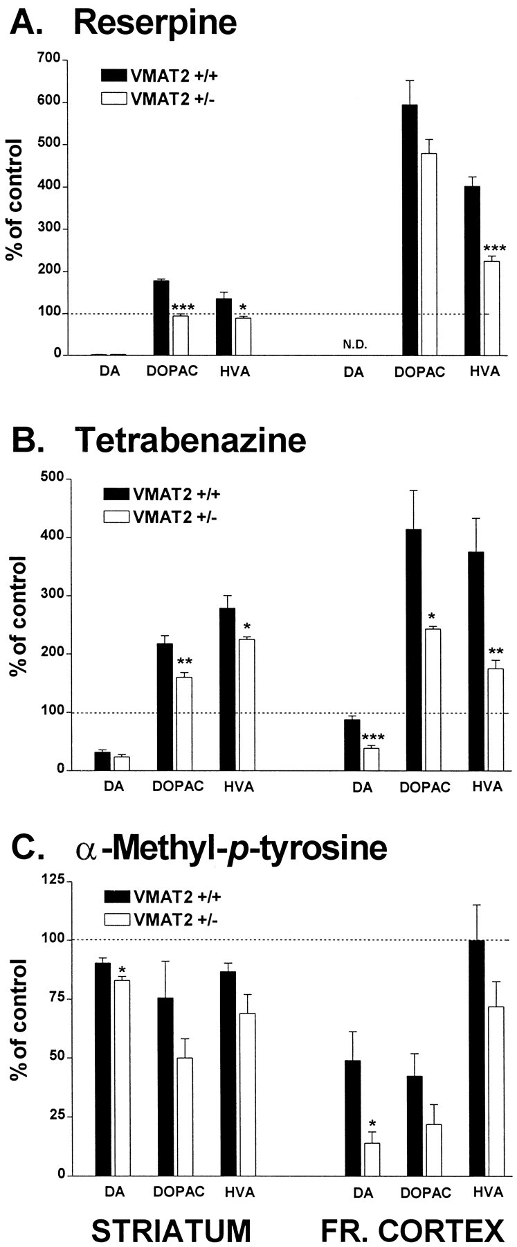
Fig. 1.

Effect of reserpine (A), tetrabenazine (B), and α-methyl-p-tyrosine (C) on DA and metabolite content in striatum and frontal cortex. The data are presented as percentages of DA, DOPAC, and HVA levels in saline-treated controls, respectively, which were as follows: striatum, 20.7 ± 2.3, 1.3 ± 0.05, and 1.6 ± 0.1 ng/mg wet tissue in wild-type mice; 15.2 ± 0.4, 1.7 ± 0.1, and 1.4 ± 0.05 ng/mg wet tissue in VMAT2 +/− mice; frontal cortex, 0.05 ± 0.03, 0.02 ± 0.01, and 0.08 ± 0.01 ng/mg wet tissue in wild-type mice; 0.06 ± 0.005, 0.03 ± 0.04, and 0.1 ± 0.005 ng/mg wet tissue in VMAT2 +/− mice. Animals treated with reserpine or tetrabenazine (5 mg/kg, i.p) were killed 6 and 1 hr, respectively, after injection. Animals treated with αMPT (250 mg/kg) were killed 1 hr after administration. Dopamine and metabolite levels were determined using HPLC-EC as described in Materials and Methods. Values represent the mean ± SEM of four to five independent determinations. ***p < 0.001; **p< 0.01; and *p < 0.05 versus treated wild-type animals.
