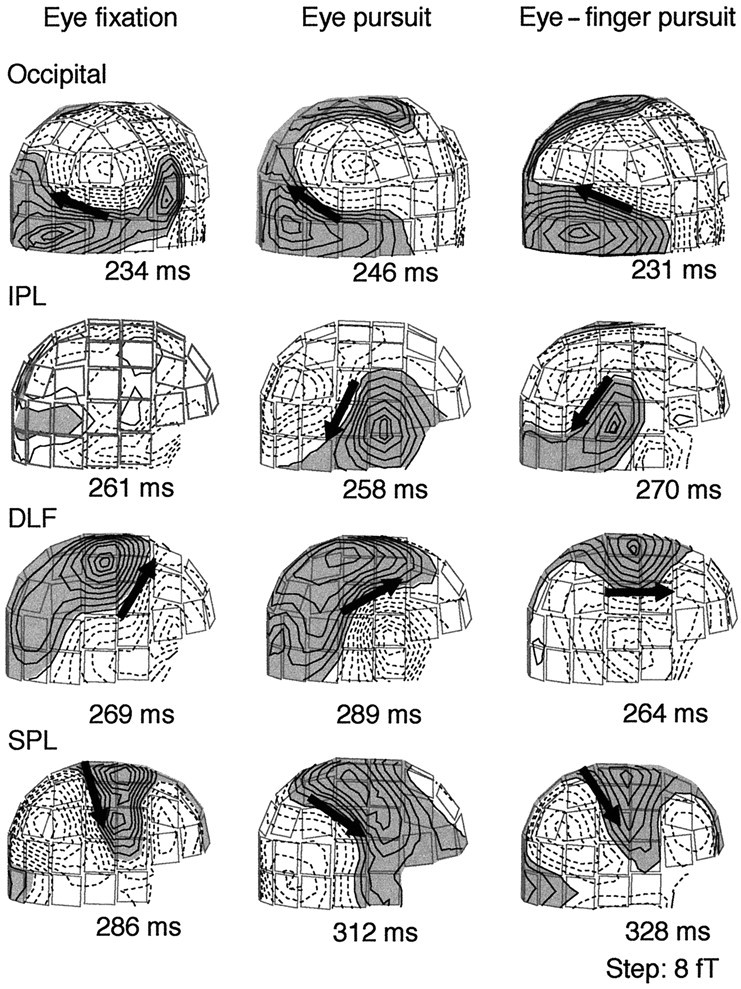
Fig. 5.

Magnetic field patterns of subject 4 during the eye fixation (left), eye pursuit (middle), and eye–finger pursuit tasks (right). The magnetic isocontour lines are separated by 8 fT. Shaded areas with solid linesillustrate the magnetic flux emerging from the head, and the areas withdotted lines illustrate the flux into the head.Arrows illustrate the locations and directions of the ECDs for dipolar field patterns; note that no ECD fulfilling the criteria was found in the IPL region during the eye fixation task.
