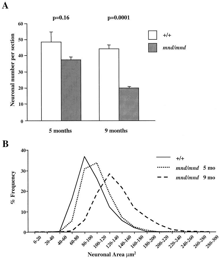
Fig. 5.
Number and size of detectable parvalbumin-stained interneurons in the entorhinal cortex of mnd/mnd mice.A, Histogram of Abercrombie-corrected number of detectable parvalbumin-positive interneurons per section of entorhinal cortex. Fewer parvalbumin-positive neurons were detected inmnd/mnd versus control (+/+) animals at 5 and 9 months, although this reduction in neuronal number was only significant in agedmnd/mnd. B, Plot of cell size distribution revealed hypertrophy of remaining parvalbumin-positive interneurons in mnd/mnd mice that was more pronounced in aged mnd/mnd.

