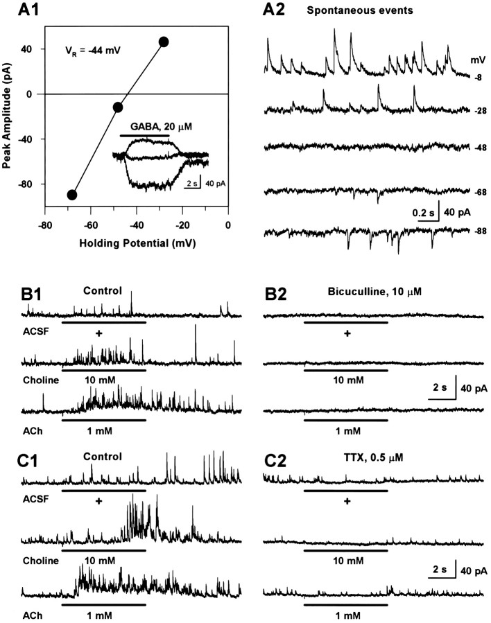
Fig. 4.
Nicotinic agonists trigger GABAergic PSCs in interneurons in an action potential-dependent manner.A1, Plot of the membrane potential versus peak amplitude of GABA-evoked currents. Data points are from the traces shown inA2, inset. Spontaneous PSCs recorded from another interneuron at different membrane potentials. Note that both spontaneous PSCs and GABA-evoked whole-cell currents reversed at approximately −44 mV. B, Recording of PSCs spontaneously occurring or evoked by nicotinic agonists in an interneuron at +2 mV. Traces on the left column(B1) were obtained under control condition, and those on the right (B2) were obtained 3–9 min after exposure of the neuron to bicuculline. C, Panel of traces showing spontaneous and agonist-evoked (6 sec pulse) PSCs under control (C1) and 3–7 min after exposure to TTX (C2). Data were obtained from a single interneuron at +2 mV. All experiments with PSCs were performed using a pipette solution that contained Cs-methanesulfonate as the main anion and QX-314 (5 mm).

