
Fig. 9.
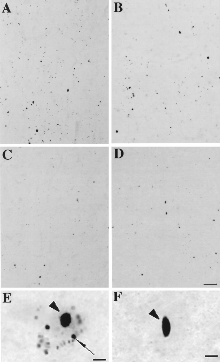
Ubiquitination of EM48 aggregates. Adjacent sections through layers V/VI of HD cerebral cortex immunolabeled with EM48 (A, B) and ubiquitin (C, D). In grade 1, the density of EM48-immunostained aggregates is three times that of ubiquitin-labeled aggregates (A, C). In grade 4, however, the densities are more similar (B, D). At higher magnification, it is evident that small perikaryal aggregates are stained by (E) EM48 but not by (F) anti-ubiquitin antibody. Scale bars:A–D, 70 μm; E, F, 10 μm.