
Fig. 5.
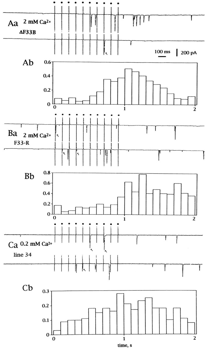
Release of transmitter during and after tetanic stimulation. Ten pulses at 10 Hz were delivered while the muscle cell was voltage-clamped at −60 mV. Aa, Current traces on a paper recorder during and after stimulation recorded in ann-sybΔF33B embryo. The external solution contained 2 mm Ca2+ and 4 mm Mg2+. Each stimulus was given at adot indicated at the top. Downward deflections indicate inward currents. Ab, The number of events per 100 msec of tetanic stimulation is plotted on the ordinate. A total of 76 sets of 10 tetanic stimuli were delivered. Timing is aligned to the top traces. Ba, Current traces on a paper recorder during and after tetanic stimulation recorded in an n-sybF33-R.Bb, Number of events per 100 msec of tetanic stimulation. A total of 59 sets of 10 stimuli were delivered. The external solution contained 2 mm Ca2+and 4 mm Mg2+. Ca, Current traces on a paper recorder during and after stimulation recorded in a line 34 larva. The external solution contained 0.2 mm Ca2+ and 5.8 mmMg2+. Arrows indicate evoked synchronized synaptic currents. Cb, Number of events per 100 msec of tetanic stimulation. A total of 119 sets of 10 tetanic stimuli were delivered.