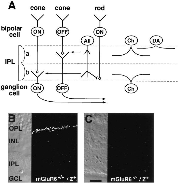
Fig. 1.
Schematic drawing of retinal cells and immunostaining of mGluR6. A, The distribution and dendritic and axonal stratifications of retinal cells. Cone ON bipolar cells make synaptic contacts with ON RGCs in sublamina b, whereas cone OFF bipolar cells form synaptic connections with OFF RGCs in sublamina a. Rod bipolar cells extend their axons to the innermost part of sublamina b, where they make synaptic contacts with AII amacrine cells (AII). AII amacrine cells form gap junctions onto cone ON bipolar cells and also project inhibitory outputs onto cone OFF bipolar cells and OFF RGCs. a, Sublamina a;b, sublamina b; Ch, cholinergic amacrine cell; DA, dopaminergic amacrine cell. B,C, Immunostaining of mGluR6 in transverse retinal sections of mGluR6+/+/lacZ+and mGluR6−/−/lacZ+mice. Punctate and intense mGluR6 immunoreactivity observed in the OPL of mGluR6+/+/lacZ+mice was absent in mGluR6−/−/lacZ+mice. Scale bar, 20 μm.

