
Fig. 2.
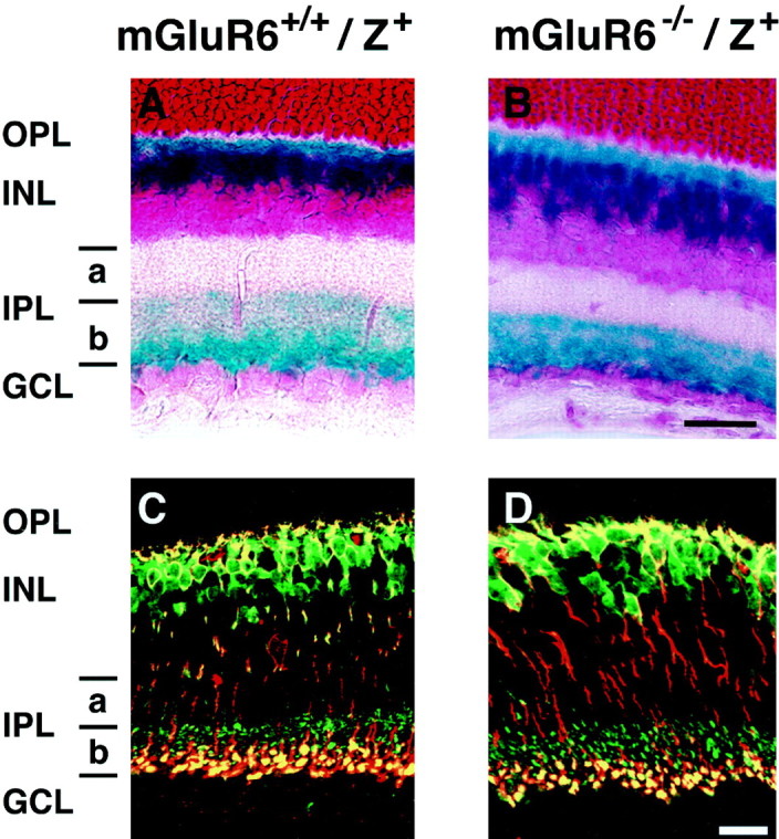
X-gal staining and double immunostaining of bipolar cells in transverse retinal sections of mGluR6+/+/lacZ+and mGluR6−/−/lacZ+mice. A, B, X-gal staining shows the localization of dendrites, somata, and axon terminals of rod and cone ON bipolar cells. C, D, The segregation of axon terminals of rod and cone ON bipolar cells is visible by double immunostaining with lacZ-Ab (green) and PKC-mAb (red);lacZ-positive/PKC-positive immunostaining at the inner half of sublamina b (yellow) andlacZ-positive/PKC-negative immunostaining at the outer half of sublamina b (green) represent axon terminals of rod bipolar cells and cone ON bipolar cells, respectively. Scale bars, 20 μm.