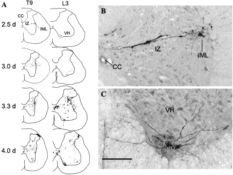
Fig. 1.
PRV immunoreactivity in the spinal cord after injection into the lordosis-producing muscles. A, Representative drawings of the morphology and distribution of PRV-immunoreactive neurons in the thoracic and lumbar spinal cord 2.5, 3.0, 3.3, and 4.0 d after PRV injection. At each time point, drawings are from the same animal. B, C, Representative PRV-immunoreactive neurons in the intermediate gray and IML (B) and the ventral horn (C) in sections taken from spinal segments T9 and L2, respectively, 2.5 d after PRV injection into the lordosis-producing muscles. Scale bar, 200 μm. CC, Central canal; IZ, intermediate zone;VH, ventral horn.

