
Fig. 8.
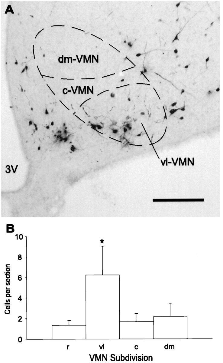
A, Representative micrograph of PRV-immunoreactive neurons in the mediobasal hypothalamus 4.0 d after PRV injection into the lordosis-producing muscles. Neurons in the VMN are predominantly in the vl-VMN. 3V, Third ventricle. Scale bar, 200 μm. B, Cell counts taken from the rostral (r), ventrolateral (vl), central (c), and dorsomedial (dm) subdivisions of the VMN 4.0 d after PRV injection into the lordosis-producing muscles. Data were collected from 10–13 sections per animal, and values are shown as mean ± SEM (n = 7). *p < 0.05.