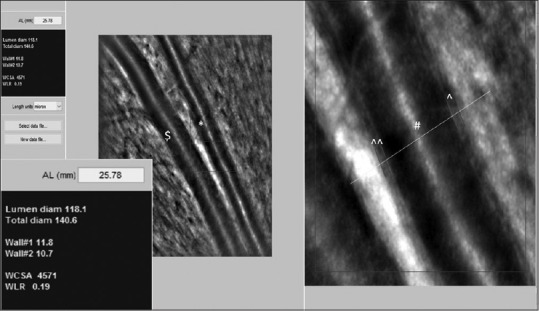
Figure 1.

Retinal arteriole and venule taken by AO-backed retinal camera. Analysis of the superotemporal arteriole parameters of the 23-year-old healthy female patient: A point on the vessel is manually selected for analysis, the rest is performed automatically by a semi-automated software AOdetect Artery 2.0b13 (Imagine Eyes, Orsay, France). Automatic detection of the vessel walls is done by detection of a rapid contrast change in the intensity across the Vernier line. The software gives the results in a side window. Inset: Magnified view of the side window with the measured six parameters. Scale bar = 100 μm ($: retinal venule; *: retinal arteriole; ^: W1; ^^: W2; #: lumen of retinal arteriole)
