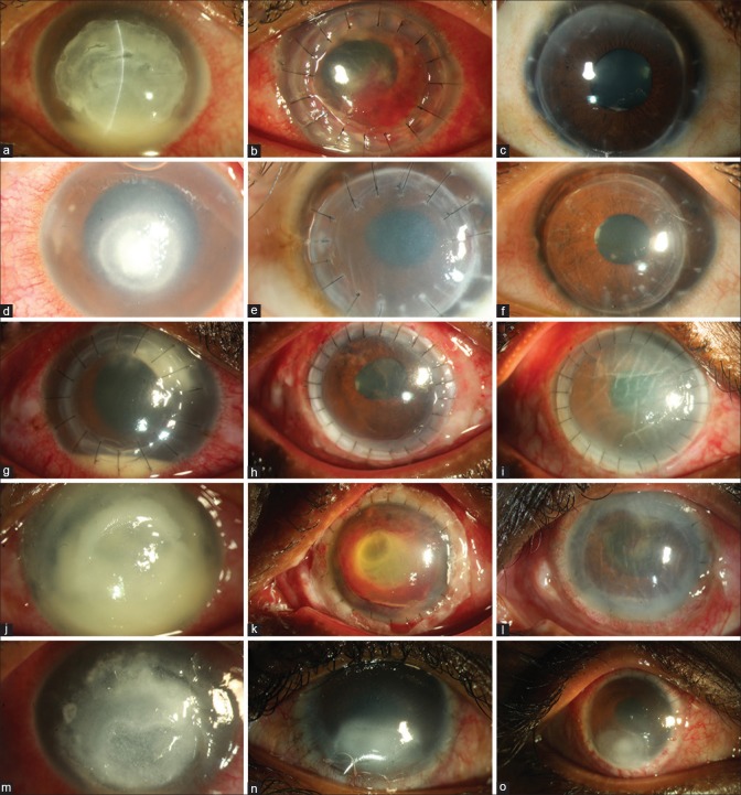
Figure 1.
Representative images of five patients: Pt 1 (a-c) had 8.5 × 8.5 mm infiltrate, 5 years post ThPK, the graft was clear, BCVA 20/20; Pt 2 (d-f) had a nonhealing fungal infection, underwent ThPK which developed a rejection episode at 10 days. Endothelial keratoplasty restored BCVA of 20/25; Pt 3 (g-i) had a recurrence, managed by repeat ThPK. The graft failed due to acute rejection at 14th day; Pt 4 (j-l) had total corneal involvement. At 5 weeks, the graft is edematous with disorganized anterior segment; Pt 5 (m-o) had 9.5 × 10 mm infiltrate (a) and underwent large ThPK. Postoperatively the graft had persistent epithelial defect that healed after tarsorrhaphy

