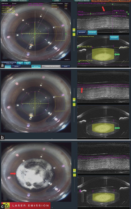
Figure 2.

Docking images. (a) Defocused laser beam for capsulotomy (red arrow) and lens fragmentation (blue arrow). (b) Laser focused on anterior lens capsule (red arrow) and lens substance (blue arrow). (c) Docking image showing entrapment of cavitation bubbles (arrow) under the ICL
