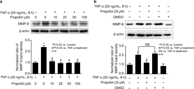
Fig. 2.
Propofol could inhibit TNF-α-induced MMP-9 expression in hCMEC/D3 cells. a The cells were pretreated with different concentrations of propofol (10, 25, 50, 100 μM) for 2 h, followed by TNF-α treatment (25 ng/mL, 8 h). The upper panel shows the protein expression of MMP-9 in hCMEC/D3 cells. The lower panel is the normalized ratio of optical densities. b DMSO served as a solvent for propofol. The upper panel shows the protein expression of MMP-9 in hCMEC/D3 cells treated with TNF-α, propofol and DMSO (0.01%). The lower panel shows the normalized ratios of optical densities, as shown by histogram. β-Actin served as an internal loading control. Data were repressed as the mean ± SD. *P < 0.05, compared with the control group. #P < 0.05, compared with TNF-α treatment group. NS = no significance. Each assay was performed in five replicates

