Fig. 5.
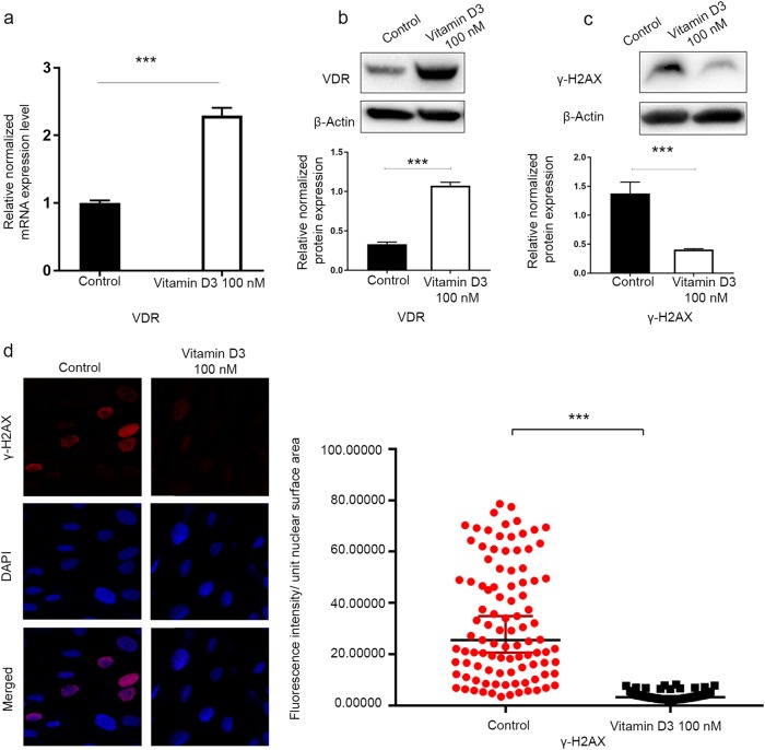
The 1, 25 dihyroxyvitamin D3 (vitamin D3) induced VDR expression and a decreased DNA damage load in HuLM cells. HuLM cells were treated with 100 nM of vitamin D3 for 3 d. Ethanol vehicle was added to the control cells. a Real-time PCR analysis of the mRNA level of the VDR gene was performed. The mRNA levels were normalized to 18 S rRNA, and normalized values were used to generate the graph. Data are presented as the mean ± SEM of triplicate measurements. Cell lysates were analyzed by Western blot analysis using (b) anti-VDR antibody and (c) anti-γ-H2AX. The intensity of each protein signal was quantified and normalized to the corresponding β-actin and presented in the graph as the mean ± SD. d HuLM cells (8 × 10⁴) were seeded on sterile glass coverslips in 6-well plates and treated with 100 nM vitamin D3 for 3 d. Ethanol vehicle was added to the control cells. γ-H2AX foci reflecting DNA DSB were assessed using immunofluorescence staining and confocal laser microscopy 40x imaging. Individual data points were the γ-H2AX focus corrected fluorescence intensity per nuclear area, as measured by ImageJ, and lines represent the median ± 95% CI. ***P < 0.001. All experiments were repeated twice

