Fig. 6.
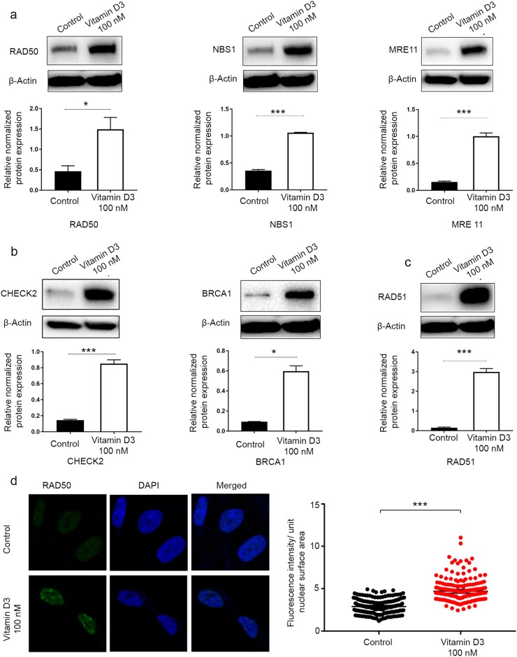
The 1, 25 dihyroxyvitamin D3 (vitamin D3) induced DNA repair capacity in HuLM cells. HuLM cells were treated with 100 nM of vitamin D3 for 3 d. Ethanol vehicle was added to the control cells. Cell lysates were analyzed by Western blot analysis to measure the protein expression of DNA double-strand breaks (DSB) repair-related markers, including homologous recombination DNA DSB (a) sensors (RAD50, NBS1, MRE11), (b) mediators and effectors (CHECK2, BRCA1), and (c) DSB binding (RAD51). The intensity of each protein signal was quantified and normalized to the corresponding β-actin and presented in the graph as the mean ± SD. d HuLM cells (8 × 10⁴) were seeded over sterile glass coverslips in six-well plates and treated with 100 nM vitamin D3 for 3 d. Ethanol vehicle was added to the control cells. RAD50 foci were assessed using immunofluorescence staining and confocal laser microscopy 40x imaging. Individual data points were the RAD50 focus-corrected fluorescence intensity per nuclear area, as measured by ImageJ, and lines represent medians ± 95% CI. *P < 0.05, ***P < 0.001. All experiments were repeated twice

