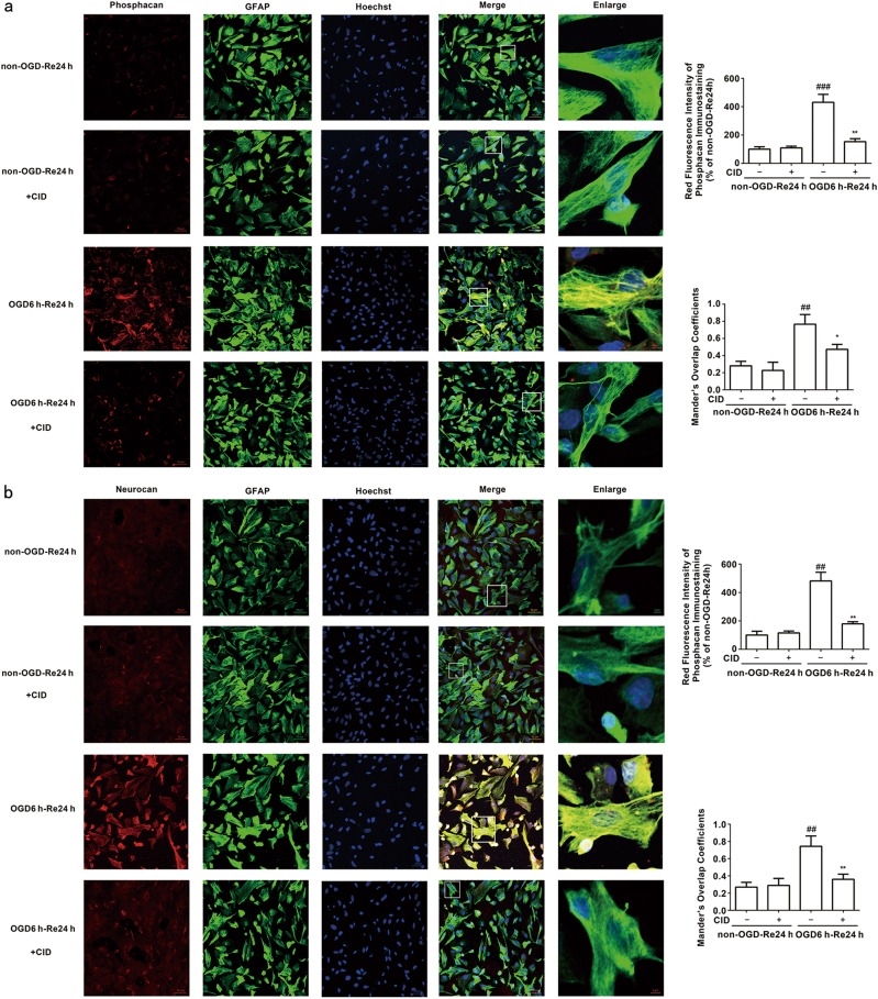
Fig. 3.
Rab7 receptor antagonist inhibits the immunoreactivity of Phosphacan and Neurocan after OGD/Re-24h. a Representative images of Phosphacan, GFAP, and Hoechst staining in astrocytes after OGD for 6 h and reoxygenaration for 24 h (Phosphacan: red; GFAP: green; Hoechst: blue). Quantification of red fluorescence intensity of Phosphacan immunostaining. Mander’s overlap coefficients demonstrated the co-localization between phosphacan and GFAP. Mean ± SD. n = 6. ##P < 0.01 vs non-OGD-Re24h group; **P < 0.01, *P < 0.05 vs. OGD6h-Re24h group. b Representative images of Neurocan, GFAP, and Hoechst staining in astrocytes after OGD for 6 h and reoxygenaration for 24 h (neurocan: red; GFAP: green; Hoechst: blue). Quantification of red fluorescence intensity of Neurocan immunostaining. Mander’s overlap coefficients demonstrated the co-localization between Neurocan and GFAP. Mean ± SD. n = 6. ##P < 0.01 vs. non-OGD-Re24 h group; **P < 0.01 vs. OGD6h-Re24h group. Statistical analysis was carried out with a one-way ANOVA followed by a post-hoc Tukey test

