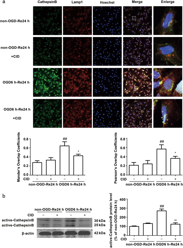
Fig. 8.
Rab7 receptor antagonist inhibits release of cathepsin B from the lysosomes into the cytoplasm after OGD/Re, and inhibits the activation of cathepsin B after OGD/Re. a Representative images of cathepsin B, Lamp1, and Hoechst staining in astrocytes after OGD for 6 h and reoxygenaration for 24 h (cathepsin B: green; Lamp1: red; Hoechst: blue). Mander’s overlap coefficients and Pearson’s overlap coefficients demonstrated the co-localization between cathepsin B and Lamp1. Mean ± SD. n = 6. ##P < 0.01 vs. non-OGD-Re24 h group; *P < 0.05 vs. OGD6h-Re24h group. b Image of active cathepsin B at OGD 6 h and reoxygenaration for 24 h with Western blotting analysis. Columns represent quantitative analysis of immunoblots. β-actin protein was used as a loading control. Mean ± SD. n = 3. ##P < 0.01 vs. non-OGD-Re24h group; **P < 0.01 vs. OGD6h-Re24h group. Statistical analysis was carried out with a one-way ANOVA followed by a post-hoc Tukey test

