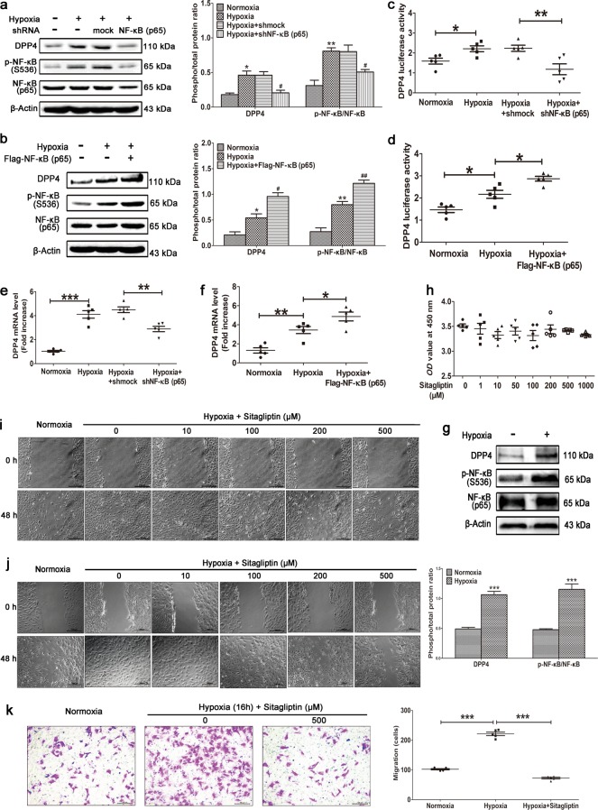
Fig. 5.
Hypoxia-induced activation of NF-κB promotes DPP4 expression. NF-κB (p65) was (a) knocked down or (b) overexpressed in primary PASMCs cultured under hypoxic conditions. Western blotting was performed with the indicated antibodies. All phospho-protein levels were measured by densitometry and normalized to that of total protein (n = 5). NF-κB (p65) was (c) knocked down or (d) overexpressed in HEK293T cells transfected with the DPP4 luciferase plasmid and cultured under hypoxic conditions. DPP4 luciferase activity was analyzed (n = 5). NF-κB (p65) was (e) knocked down or (f) overexpressed in primary PASMCs cultured under hypoxic conditions. Quantitative RT-PCR was performed to examine the mRNA level of DPP4 (n = 5). g Primary cultured human PASMCs were stimulated by hypoxia, and Western blotting was performed with the indicated antibodies. phospho-p65 levels were measured by densitometry and normalized to that of total protein (n = 5). h Cytotoxicity assay using primary cultured PASMCs stimulated with various concentrations of sitagliptin (n = 5). Primary rat PASMCs (i) and primary human PASMCs (j) were cultured under hypoxic conditions. When almost fully confluent, PASMCs were wounded by scraping and treated with the indicated concentration of sitagliptin. Images were acquired at 0 and 48 h (n = 6). k Human PASMCs were cultured in a transwell upper chamber and stimulated with hypoxia and sitagliptin. The migrated cells in the lower chamber were stained with a 1% crystal violet solution after 15 h and photographed for statistical analysis (n = 5). The data are expressed as the means ± standard errors of the mean. */#P < 0.05, **/##P < 0.01, ***P < 0.001

