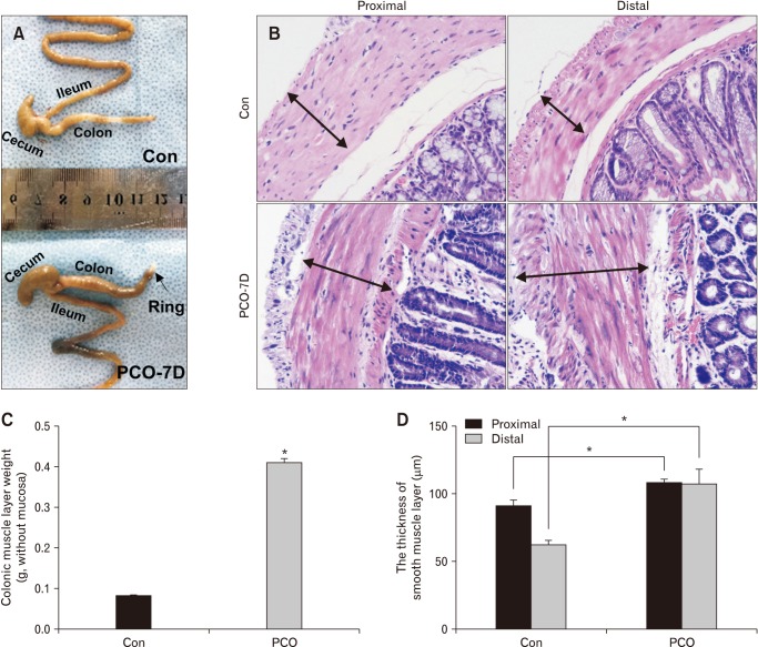
Figure 1.
Surgically induced partial colon obstruction (PCO) in a mouse model. (A) Pictures showing the anatomic changes in control (Con) and PCO mice. (B, D) Comparison of the thickness of the smooth muscle layer between control and PCO mice by H&E staining (×200 magnification) and statistical analysis (*P < 0.05, n = 7). (C) Summarized data showing the colonic muscle layer weight in control and PCO mice (*P < 0.05, n = 7).

