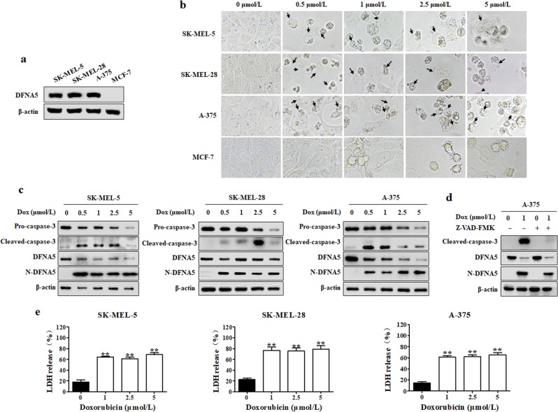
Fig. 1.
Doxorubicin induces pyroptosis in melanoma cells. a Differential expression levels in DFNA5 in SK-MEL-5, SK-MEL-28, A-375, and MCF-7 cells. The DFNA5 levels in SK-MEL-5, SK-MEL-28, A-375, and MCF-7 cells were measured by Western blot. β-actin was used as the loading control. b SK-MEL-5, SK-MEL-28, A-375, or MCF-7 cells were treated with a series of doxorubicin (Dox) concentrations for 24 h. Pyroptotic morphologies were observed at ×400 magnification under an inverted fluorescence microscope. Arrowheads indicate pyroptotic cells. c SK-MEL-5, SK-MEL-28, and A-375 cells were treated with a series of doxorubicin (Dox) concentrations for 24 h. The levels of DFNA5, N-DFNA5, caspase-3, and cleaved caspase-3 were measured by Western blot. β-actin was used as the loading control. d A-375 cells were pretreated with 10 μmol/L Z-VAD-FMK for 1 h, followed by doxorubicin treatment for 24 h. After treatment, the cleaved caspase-3 and N-DFNA5 levels were measured by Western blot. β-actin was used as the loading control. e At the end of treatment, the LDH levels were measured. Each bar represents the mean ± SD of triplicate measurements from one of three identical experiments. *P < 0.05, **P < 0.01 vs. the control group

