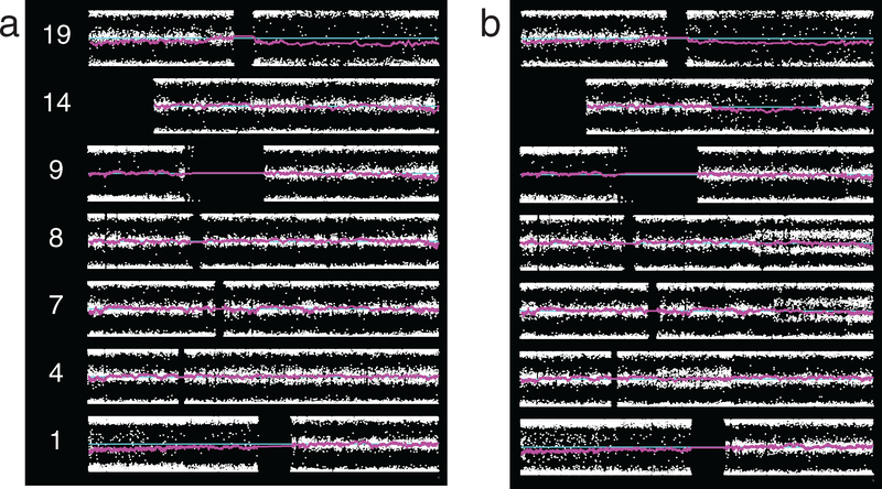
Figure 2.
Representative SNP microarray data plots from ODG, two patients. Plots show chromosomes of interest, p-arm on left, with BAF at each SNP location (white dots), smoothed LRR (magenta), and neutral LRR position (cyan). (a) Grade II tumor, case 2_40. Whole arm broadly split BAF is seen on 1p and 19q. LRR shows this to be deletion. In addition to 1p/19q, this case demonstrates thinly split BAF along most of 4q, karyotypic change indeterminate, and broadly split BAF along most of 9p due to copy-neutral change. (b) Grade III tumor, case 3_11. Beyond 1p/19q, this case demonstrates broad splitting on the whole of 9p due to copy-neutral LOH, segmental thin splitting on 4q, 7q, and 8q due to indeterminate karyotypic change, and and broad splitting on a segment of 14q due to deletion.

