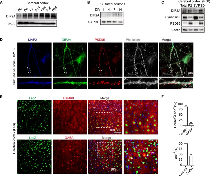
Fig 1. DIP2A is localized to dendritic spines in excitatory neurons.
(A) Western blotting showing the level of DIP2A in cerebral cortex constantly increased during postnatal development. (B) Level of DIP2A in cultured neurons. (C) Western blot showing DIP2A in the cortical PSD composition (lane 4). Total, total homogenate; P2, crude synaptosomes; SV, crude synaptic vesicle fraction. (D) Representative image showing the distribution of MAP2 (blue), DIP2A (green), and PSD95 (red) in cultured neurons. Dashed white boxes represent the enlarged view. (E) Dip2a β-galactosidase (LacZ) reporter mice (Dip2alacZ/+) were used to display endogenous DIP2A-expressed cell types. CaMKII and GABA were stained in cortical sections of Dip2alacZ/+ mice to label excitatory and inhibitory neurons with antibodies, respectively. Arrowhead, GABA immunoreactive neurons; arrow, GABA and LacZ double staining neurons. (F) Relative quantification of LacZ-, CaMKII-, and GABA-positive cells. The underlying data for this figure can be found in S1 Data. CaMKII, Ca2+/calmodulin-dependent protein kinase II; DIP2A, disconnected-interacting protein homolog 2 A; DIV, day in vitro; GAPDH, glyceraldehyde-3-phosphate dehydrogenase; LacZ, Dip2a β-galactosidase; MAP2, microtubule-associated protein 2; P, postnatal day; PSD, postsynaptic density; P2, crude synaptosomes; SV, crude synaptic vesicle fraction.

