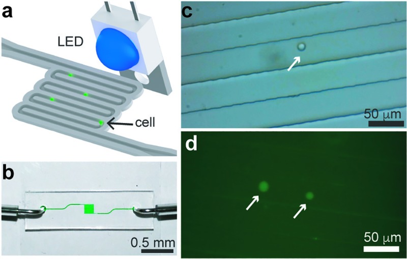
Fig 7. Tracking cells in a microfluidic channel.

(a) Schematic of the microfluidic device in which cells stained with a fluorescent dye are flowed in a microfluidic device. (b) Photograph of the single-channel microfluidic device. (c) Bright-field image of a single THP-1 cell flowing at a speed of ~150 μm/s in a 40-μm wide channel. (d) Fluorescence micrographs of two cells flowing inside channel with a 100 ms exposure time. White arrows point to cells.
