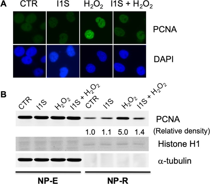
Fig 4. The effects of H2O2 and PCNA-I1S on PCNA association with chromatin.
A. PC-3 cells (2 x 10⁴/well) were plated into chamber slides and incubated overnight. After starvation in serum-free medium for 3 days, the cells were treated with PCNA-I1S (1 uM) for 1.5 hours and/or H2O2 (100 uM) for 1 hour. The cells were lysed in a hypotonic buffer containing 0.5% NP-40, fixed in cold methanol, stained with PCNA antibody (PC10) and fluorescent secondary antibody. The cells were counterstained with DAPI to reveal cell density and nucleus. B. The cells were starved and treated as described in A. The NP-E and NP-R proteins were analyzed by immunoblotting to detect the free-form and chromatin-bound PCNA with α-tubulin and histone H1 levels as loading controls for NP-E and NP-R PCNA, respectively. The optical density of chromatin-bound PCNA was normalized to histone H1. The raw images of immunoblotting are provided as the supplementary data (S1 Raw Images).

