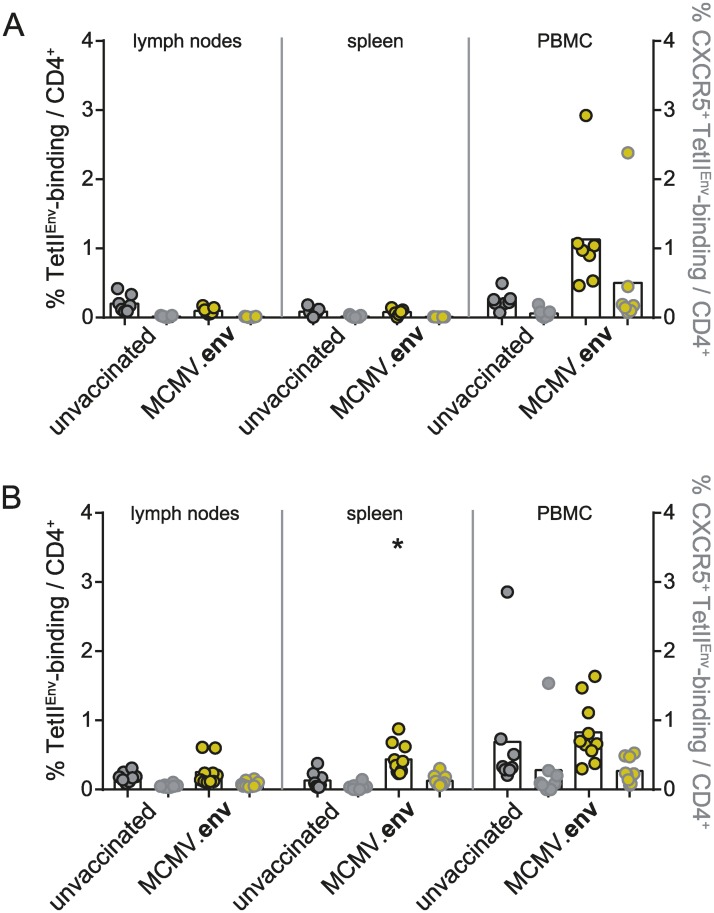
Fig 7. Distribution of F-MuLV-specific CD4+ T cells after FV challenge infection.
CB6F1 mice were immunized once with MCMV.env and infected six weeks later with 5 000 SFFU FV. Two weeks after immunization (A), or three weeks after the FV infection (B), spleen, lymph node and peripheral blood mononuclear cells were collected and subjected to MHC II tetramer staining for detection of Env123-141-specific CD4+ T cells in combination with staining for the chemokine receptor CXCR5 (grey symbols). Data were obtained in two independent experiments, each dot indicates one mouse, bars indicate mean values. Data were analysed for statistically significant differences by One Way ANOVA on Ranks with Dunn’s post test. * indicates statistically significant differences compared to the respective data from unvaccinated mice (P < 0.05).

