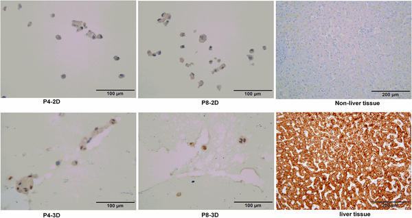
Figure 8.
Immunostaining of OTCD-PHH with hepatocyte marker antibody. Cells from 2D or 3D culture were fixed with 10% formalin overnight at 4 °C. Afterward, the cells were embedded, sliced and stained with monoclonal mouse anti-human hepatocyte antibody for 1h. Then, the slides were washed and incubated with biotin-conjugated goat anti-mouse secondary antibody for 30 minute. Magnification, 40×; bar, 100 μm.

