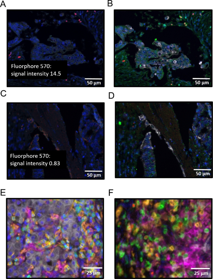
Figure 3: Successes and pitfalls of monoplex and multiplex.
(A) Monoplex assay of primary colon cancer with FOXP3 at position 3, fluorescent intensity label shown. (B) Multiplex assay of primary colon cancer with FOXP3 at position 3 and CD3 at position 2. (C) Monoplex assay of primary colon cancer with FOXP3 at position 1, fluorescent intensity label shown. (D) Multiplex assay of primary colon cancer with FOXP3 at position 1 and CD3 at position 2. (E) Multiplex assay of primary colon cancer with order of antibodies as follows: CD8 (yellow), CD3 (green), CD163 (orange), pancytokeratin (white), PD-L1 (magenta), FOXP3 (red), working DAPI (blue). (F) Multiplex assay of primary colon cancer with order of antibodies as follows: CD8 (yellow), CD3 (green), CD163 (orange), pancytokeratin (white), PD-L1 (magenta), FOXP3 (red), non-working DAPI (blue).

