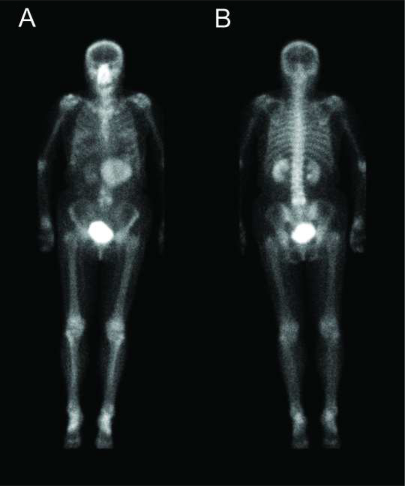
Figure 1 –
Temporal bone CT in a 75-year-old patient for mixed sensorineural and conductive hearing loss demonstrated a nonspecific sclerotic focus in the right occipital condyle. Anterior and posterior planar images from a subsequent whole body 99m-Tc-MDP bone scan were negative for suspicious osseous accumulation of radiotracer, but instead demonstrated a large discrete mass-like accumulation of extra-osseous 99m-Tc-MDP in the anterior left upper quadrant. Differential for this finding in an elderly patient included gastric lymphoma or cancer (1,2), metastatic melanoma (3,4) and heterotopic mesenteric ossification, such as from prior gastric bypass surgery (5,6). Gastric 99m-Tc-MDP uptake also has been observed in sarcoidosis (7) and diseases that cause hypercalcemia (8), including multiple myeloma (9), hyperparathyroidism (10) and vitamin D intoxication (11). Isolated splenic infarction and solitary metastasis from a mucinous ovarian or colon cancer seemed unlikely (1). A contrast-enhanced CT was obtained for further characterization.

