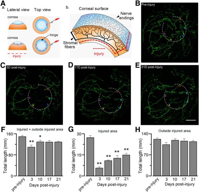
Figure 1.
In vivo confocal tracking of whole corneal innervation by TRPM8BAC-EYFP nerves. A, Schematic representation of the injury of corneal nerve fibers. Aa, Initial incision and extension of the surgical damage. Ab, Scheme of corneal innervation (showing stromal nerve bundles and corneal subbasal nerve plexus), highlighting the deep injury of the fibers. B, YFP fluorescence of intact corneal nerves (preinjury) in a TRPM8BAC-EYFP mouse in vivo. Three TRPM8BAC-EYFP mice where used for the tracking. C–E, Confocal tracking of the regeneration of axotomized corneal nerves at different time points in the same cornea: (C) 3 d after injury; (D) 17 d after injury; and (E) 21 d after injury. Light gray dotted lines indicate the axotomized area. The colored asterisks indicate different axotomized nerves at different time points. Scale bar, 500 μm. F, Total length of corneal cold sensory nerves. Statistical significance was determined by one-way ANOVA (F(4,10) = 5.878, *p = 0.0110) followed by Holm–Sidak method (preinjury vs 3 d, t(14) = 4.847, **p = 0.0038; preinjury vs 10 d, t(14) = 2.435, *p = 0.0350). G, Total length of corneal cold sensory nerves in injured area. Statistical analysis was performed using one-way ANOVA (F(4,10) = 38.727, ***p < 0.0010) followed by Holm–Sidak method (preinjury vs 3 d, t(14) = 11.857, **p = 0.0038; preinjury vs 10 d, t(14) = 8.960, **p = 0.0029; preinjury vs 17 d, t(14) = 8.014, **p = 0.0020; preinjury vs 21 d, t(14) = 6.747, *p = 0.0010). H, Total length of corneal cold sensory nerves in intact areas. Statistical analysis was assessed with a one-way ANOVA (F(4,10) = 1.288, n.s. p = 0.3380). (**p < 0.05; **p < 0.01).

