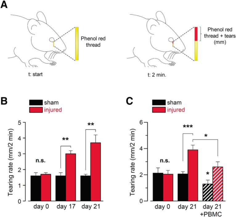
Figure 7.

Changes in basal tear secretion rate in response to corneal nerve damage. A, Schematic representation of the measurement of tearing rate by phenol red thread test in mice. B, Basal tearing rate, expressed as the mean wetted length of the phenol red-treated thread in mm after 2 min, measured in anesthetized mice (n = 16 in each condition; t(30) = 0.0425, n.s. p = 0.9664 (day 0), t(30) = 3.473, **p = 0.0016 (day 17), unpaired t test, and t(18) = 3.846, **p = 0.0012 (day 21), unpaired t test with Welch's correction). C, Basal tearing rate, expressed as the mean wetted length of the phenol red-treated thread in mm after 2 min, measured in a different set of anesthetized mice. At day 21, the graph shows the mean tearing rate in sham (n = 9) and injured (n = 10) animals, before (solid bars) and after (dashed bars) the application of 20 μm PBMC on the corneal surface. (t(17) = 0.2452, n.s. p = 0.8092 (day 0), unpaired t test, and t(12) = 4.753, ***p = 0.0005 (day 21), unpaired t test with Welch's correction; t(8) = 2.485, *p = 0.0378 (day 21, sham mice before and after PBMC), and t(9) = 2.899, *p = 0.0176 (day 21, injured group before and after PBMC), paired t test).
