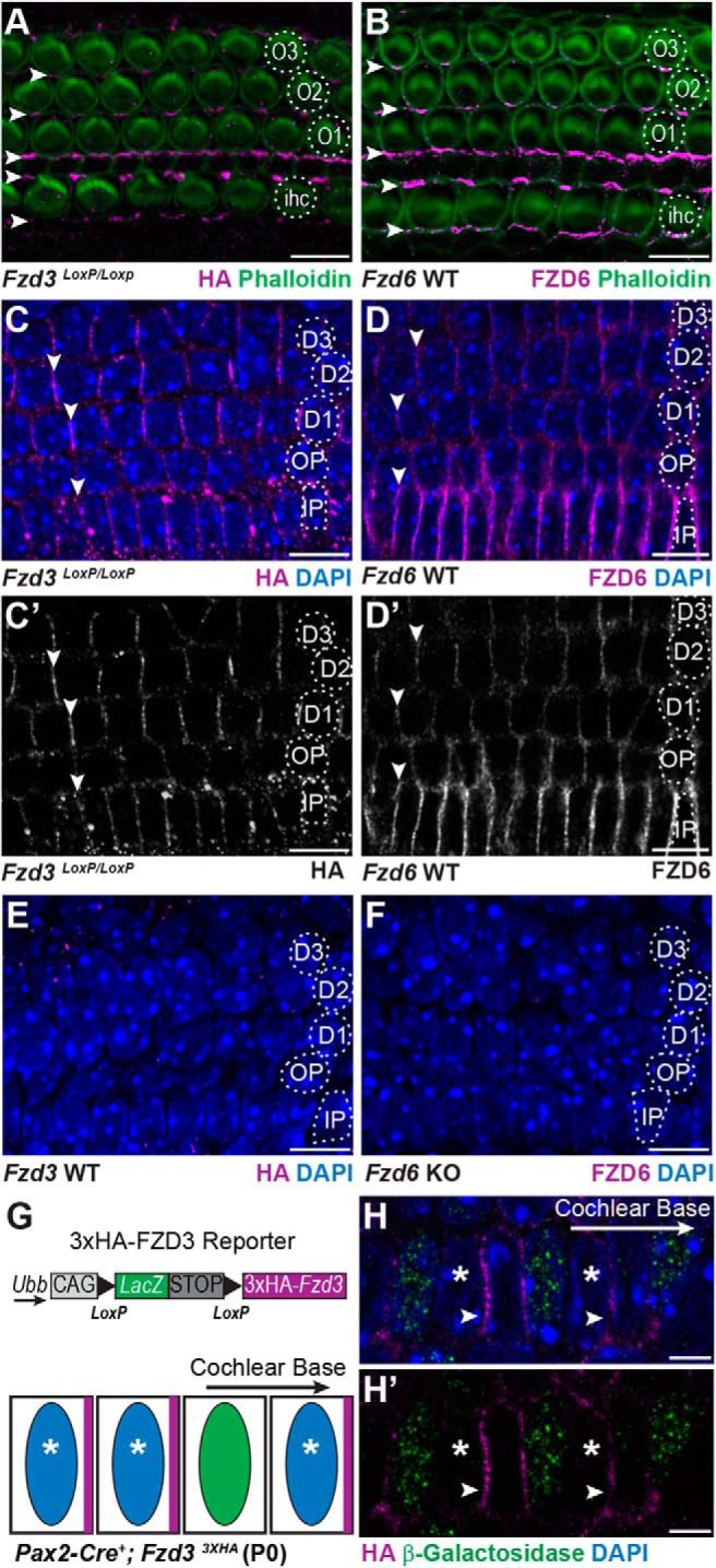
Figure 2.

FZD3 and FZD6 are asymmetrically distributed along the basolateral sides of cochlear-supporting cells. A, HA immunofluorescence at P0 is enriched at hair cell to supporting cell junctions on the apical surface of the organ of Corti in a Fzd3 CKO mouse in which endogenous FZD3 contains an HA epitope tag (arrowheads). B, FZD6 protein is similarly enriched at hair cell to supporting cell junctions (arrowheads). C, C′, FZD3-HA and D, D′, FZD6 are also enriched along the basolateral surfaces of supporting cells at supporting cell to supporting cell junctions (arrowheads). E, Specificity of the HA antibody demonstrated by immunolabeling the basolateral surfaces of supporting cells in WT tissue and (F) specificity of the FZD6 antibody demonstrated in Fzd6 KO tissue. G, Schematic illustration of a Cre-dependent 3XHA-Fzd3 transgenic reporter and predicted distribution of the 3XHA-FZD3 protein product (magenta) in adjacent Cre-positive (blue nuclei, asterisk) and Cre-negative (green nuclei) cells. H, H′, 3XHA-FZD3 expression and localization (arrowheads) in the Pax2-Cre+ IPCs (asterisk) on the side facing the cochlear base. β-galactosidase immunolabel (green) marks Cre-negative cells, which do not express the FZD3 reporter. IHCs, first (O1), second (O2), and third (O3) row OHCs, IPCs, OPCs, first (D1), second (D2), and third (D3) row Deiters' cells. All images are of P0 tissue. Scale bars: A–F, 10 μm; H, 5 μm.
