Figure 4.
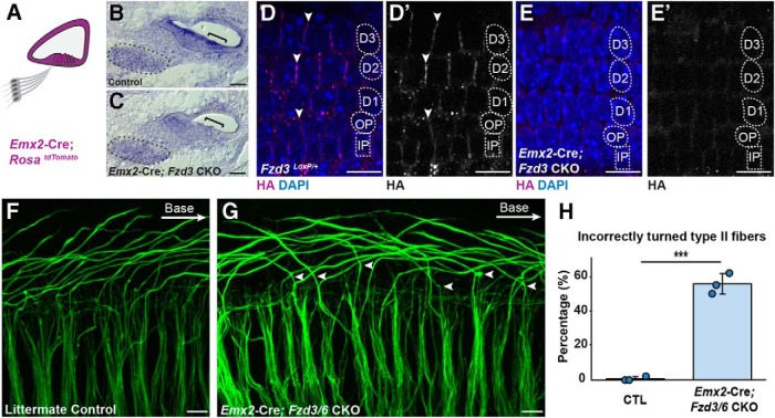
Type II turning is regulated by nonautonomous Fzd3/Fzd6 function. A, Schematic diagram showing the pattern of Emx2-Cre-mediated recombination in the cochlear duct. B, C, Conventional ISH showing that the reduction of Fzd3 mRNA at E16.5 in the organ of Corti (bracket) but not the SGN (outlined) from Emx2-Cre;Fzd3 CKOs. D, D′, E, E′, FZD3-HA expression is also lost from the basolateral junctions between neighboring supporting cells in Emx2-Cre;Fzd3 CKOs compared with littermate controls. F, G, NF200 immunolabeling of Type II SGN peripheral axons in Emx2-Cre;Fzd3 CKO;Fzd6 KOs (Emx2-Cre;Fzd3/6 CKO) reveals multiple turning errors (arrowheads) compared with littermate controls in the basal turn of the E18.5 cochlea. H, Average frequency of incorrectly turned Type II fibers projecting toward the cochlear apex. Data are mean ± SD with overlying circles reporting to the frequency of turning errors for individual mice. A single cochlea was analyzed from each animal: Control (n = 3, 127 axons), CKOs (n = 3, 142 axons). ***p ≤ 0.001, CKOs versus littermate controls (Student's t test). Supporting cell nuclei are identified based upon DAPI staining of nuclei: IPCs, OPCs, first (D1), second (D2), and third (D3) row Deiters' cells. Scale bars, 10 μm.

