Figure 1.
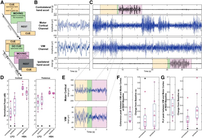
Task timeline, raw and spectral analyses. A, Task timeline of the recordings. The top timeline did not have a preparation cue. Cued cup reaching task was administered only in the top timeline format. Boxes with a dashed border represent the uncued actions the subject took during the task. B, C, E, Data collected in an ET DBS implantation surgery along with contralateral and ipsilateral hand accelerations. The subject was asked to clench his/her hand repetitively when CUED (yellow). The GO CUE event (green) represents when the subject was asked to move their hand, and the MOVING event (violet) represents when the subject moved their hand. B, Baseline/rest data were collected in a separate run. D, Normalized PSD for both contralateral and ipsilateral movement for LFO (8–30 Hz) and HBA (>40 Hz) in motor cortex (Wilcoxon signed-rank test, LFO p = 0.001 and HBA p = 0.003 for baseline vs contralateral movement, LFO p = 0.002 and HBA p = 0.413 for baseline vs ipsilateral) and VIM (Wilcoxon signed-rank test, LFO p = 0.001 and HBA p = 0.001 for baseline vs contralateral movement, LFO p = 0.002 and HBA p = 0.067 for baseline vs ipsilateral). E, Zoomed section of LFP from C during contralateral hand movement. F, Thalamocortical coherence in baseline/rest on the right, normalized coherence for both contralateral and ipsilateral movement on the left (Wilcoxon signed-rank test, p = 0.002 for baseline vs contralateral movement, p = 0.0137 for baseline vs ipsilateral). G, Thalamocortical PLV in baseline/rest on the right, normalized PLV for both contralateral and ipsilateral movement on the left (Wilcoxon signed-rank test, p = 0.001 for baseline vs contralateral movement, p = 0.003 for baseline vs ipsilateral). *p < 0.01.

