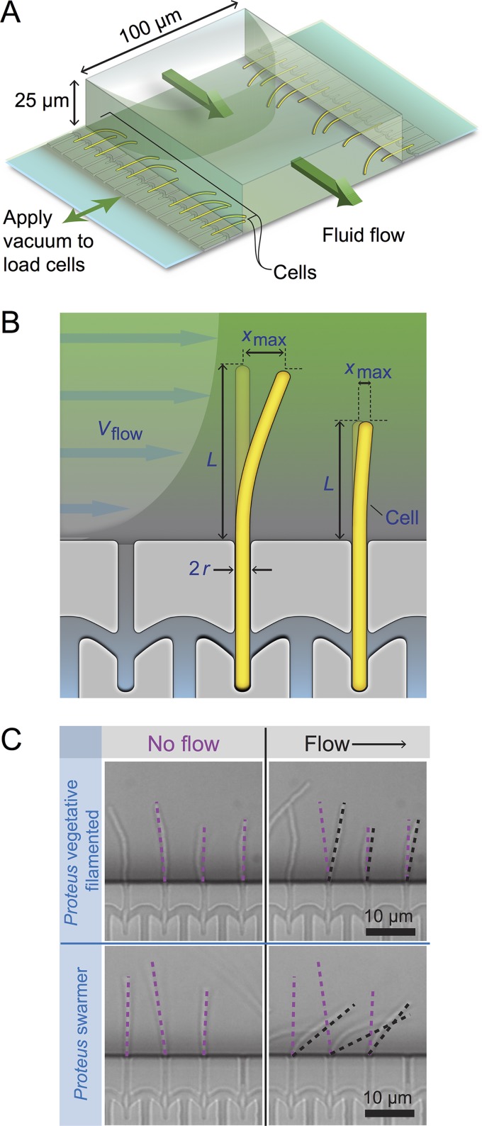
FIG 2.
Using a reloadable microfluidics-based assay to determine bacterial cell stiffness. (A) Schematic of the microfluidic channel used to apply a user-defined shear force to bend filamentous or swarmer cells. Single-sided green arrows depict the flow of fluid through the central channel; the parabolic flow profile of the fluid is shown. Double-sided green arrows indicate the vacuum chamber used to load cells into side channels and to empty the device. (B) Cartoon of a flexible bacterium (left) and a stiff bacterium (right) under conditions of flow force (Vflow). “xmax” indicates the deflection of cells in the flow. 2r = cell diameter; L = cell length in contact with the flow force. (C) Representative images of filamentous cells of P. mirabilis under no-flow (left) and flow (right) conditions (top) and P. mirabilis swarmer cells (bottom). Purple dashed lines indicate the position of a cell tip under no-flow conditions, and black dashed lines illustrate the position after flow is applied using a gravity-fed mechanism. The arrow indicates the direction of fluid flow in the channel.

