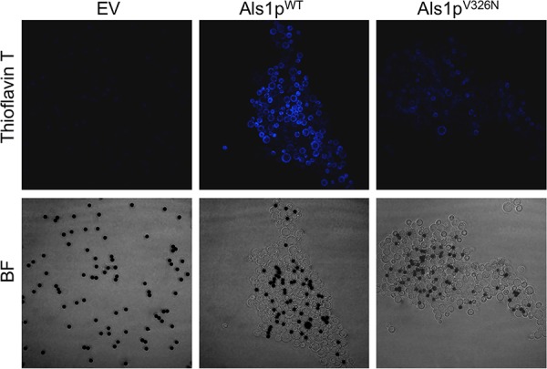
FIG 2.

Effect of V326N mutation on amyloid formation. Cell surface amyloid formation was monitored with ThT in adhesion assays with BSA-coated magnetic beads (dark spheres, 1-μm diameter). Confocal microscopy was used to examine S. cerevisiae cells containing the empty vector (EV), cells expressing Als1pWT, and cells expressing Als1pV326N. Pictures were taken at 1,000× total magnification. Overall, the aggregates were of similar size to those shown in Fig. 1C. For the purposes of fluorescence comparison, we illustrate aggregates of Als1pWT and Als1pV326N that are of similar size.
