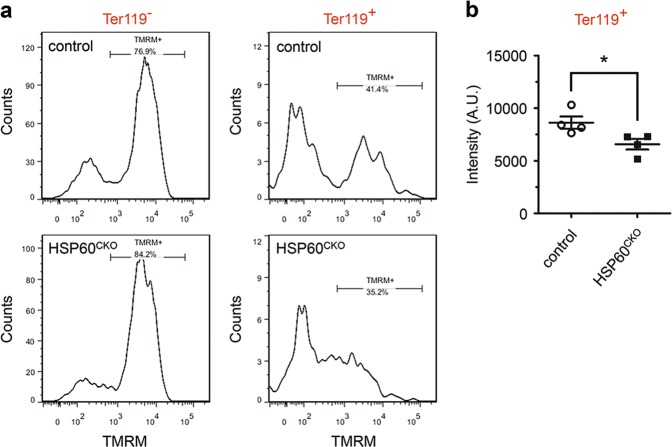
Fig. 4. Deletion of HSP60 reduced mitochondrial membrane potential in yolk sac erythrocytes.
Single cells were prepared from control and HSP60CKO yolk sacs at E8.5, and flow cytometry was performed to measure mitochondrial membrane potential using tetramethylrhodamine (TMRM) in Ter119 positive (Ter119+) and Ter119 negative (Ter119−) cells. a Distribution of TMRM fluorescence of Ter119− and Ter119+ cells in control and HSP60CKO yolk sac cells. b Statistical analysis showing reduced TMRM fluorescence in HSP60CKO Ter119+ cells. n = 4 mice per group. All data represent mean ± SEM. Significance was determined using a 2-tailed, unpaired Student’s t test. *p < 0.05 versus control

