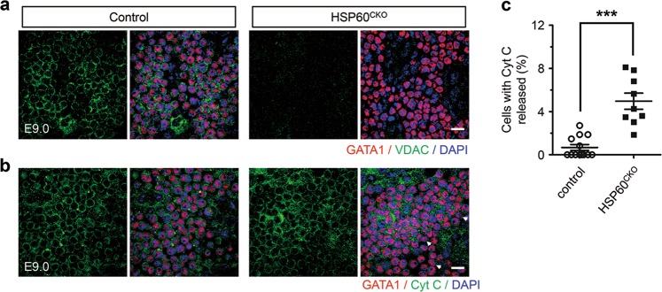
Fig. 5. HSP60 deletion affected VDAC expression and Cyt C release in yolk sac erythrocytes.
a Immunofluorescence staining of GATA1 and VDAC in control and HSP60CKO yolk sacs at E9.0. HSP60 deletion abolished VDAC expression in yolk sac erythrocytes. Data are representative of at least three independent experiments. Scale bar, 20 μm. b Immunofluorescence staining of GATA1 and Cyt C in control and mutant yolk sacs at E9.0. The GATA1 positive cells with released Cyt C were characterized with no perinuclear immunofluorescence (white arrows). Data are representative of at least three independent experiments. Scale bar, 20 μm. c Statistical analysis showing increased numbers of the cells with released Cyt C in mutant erythrocytes. n = 13 from 5 control yolk sacs and 9 from 3 HSP60CKO yolk sacs. All data represent mean ± SEM. Statistical significance was determined by Mann-Whitney U test. *p < 0.001 versus control

