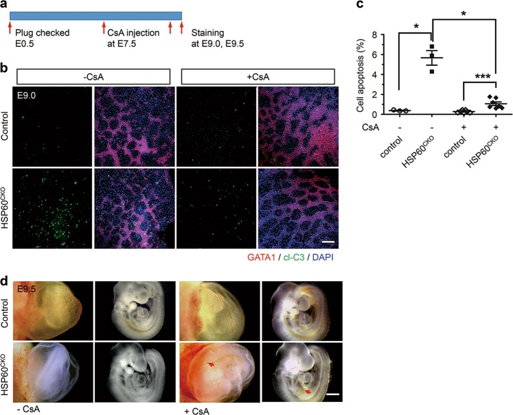
Fig. 6. Cyclosporine A reduced cell apoptosis induced by HSP60 deletion in mutant erythrocytes.
a Schematic diagram showing the procedure of the rescue experiment. Pregnant mice were intraperitoneally injected with cyclosporine A (CsA) at E7.5. The embryos were dissected at E9.0 or E9.5, and immunofluorescence staining was then performed. b Immunofluorescence staining of GATA1 and cleaved-Caspase 3 (cl-C3) in control and mutant yolk sacs treated with (+CsA) and without (−CsA) CsA at E9.0. Scale bar, 100 μm. c Statistical analysis showing that CsA treatment could significantly reduce cell apoptosis of erythrocytes in mutant yolk sacs (n = 3, −CsA; n = 7, +CsA) compared with control yolk sacs (n = 3, −CsA; n = 7, +CsA). Data represent mean ± SEM. Significance was determined using the 2-way ANOVA analysis with a Bonferroni post-hoc test. *p < 0.05, ***p < 0.001. d Morphology of control and mutant yolk sacs and embryos treated with CsA (+CsA) and without CsA (−CsA) at E9.5. Red arrows indicate vessels with red blood cells in mutant yolk sacs and embryos treated with CsA. Scale bar, 0.5 mm

